Soft tissue grafting has become an indispensable component of contemporary periodontal therapy, representing one of the most predictable and successful procedures in our clinical armamentarium. As our understanding of tissue biology and healing mechanisms continues to evolve, so too must our approach to these procedures. The integration of advanced surgical techniques, innovative materials, and precision instrumentation has transformed soft tissue grafting from a purely functional procedure to one that achieves both optimal health outcomes and superior esthetic results.
Fundamentals of Soft Tissue Grafting
Understanding the biological foundation of soft tissue grafting is paramount to achieving predictable results. The periodontal tissues exist in a delicate balance, with the gingiva serving as the primary barrier against bacterial invasion while providing the esthetic framework for the dentition. When this barrier is compromised through recession, the resulting exposure of root surfaces creates both functional and esthetic challengesthat require careful consideration.
The vascular supply of the periodontal tissues plays a critical role in graft success. The gingiva receives its blood supply from three sources: the supraperiosteal vessels, the vessels of the periodontal ligament, and the vessels emerging from the alveolar bone. Preservation of this vascular architecture during surgical procedures is essential for optimal healing and long-term stabilityof grafted tissues.
Indications for soft tissue grafting include:
-
Root coverage for esthetic concerns
-
Reduction of root sensitivity
-
Facilitation of plaque control
-
Prevention of further recession
-
Augmentation of keratinized tissue width
However, not all recession defects are suitable for grafting. Contraindications include:
-
Active periodontal disease
-
Poor oral hygiene
-
Heavy smoking
-
Systemic conditions that compromise healing
Careful patient selection remains one of the most important factors in achieving successful outcomes.
Classification of Gingival Recession
Proper classification of gingival recession defectsis fundamental to treatment planning and prognosis determination. Miller's classification system, introduced in 1985, has long served as the standard for categorizing recession defects based on the relationship between the marginal tissue recession and the mucogingival junction, as well as the presence or absence of interproximal bone and soft tissue loss.
-
Class I defects involve marginal tissue recession that does not extend to the mucogingival junction, with no periodontal loss in the interdental area. These defects offer the most favorable prognosis for complete root coverage.
-
Class II defects extend to or beyond the mucogingival junction but still have no periodontal loss interdentally. While complete root coverage is still achievable, the prognosis is slightly less favorable than Class I defects.
-
Class III and IV defects involve interproximal periodontal loss:
-
Class III shows recession extending to or beyond the mucogingival junctionwith interproximal bone or soft tissue loss.
-
Class IV presents with severe interproximal loss extending to the apical extent of the marginal tissue recession.
These defects have a guarded to poor prognosis for complete root coverage.

The more recent Cairo classification system (RT1, RT2, RT3) provides additional refinement in categorizing recession defects based on interproximal clinical attachment level, offering improved prognostic accuracy and treatment planning guidance:
-
RT1 defects: No interproximal clinical attachment loss
-
RT2 defects: Interproximal clinical attachment loss less than or equal to the buccal clinical attachment loss
-
RT3 defects: Interproximal clinical attachment loss greater than the buccal clinical attachment loss
Pre-Surgical Tissue Preparation and Contouring
Meticulous pre-surgical tissue preparation is often the difference between good and exceptional graft outcomes. The recipient site must be carefully evaluated and prepared to optimize conditions for graft integration and healing. This phase requires precision and attention to detail that extends beyond traditional periodontal instrumentation.
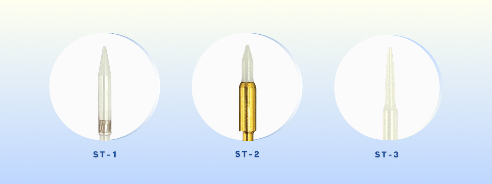
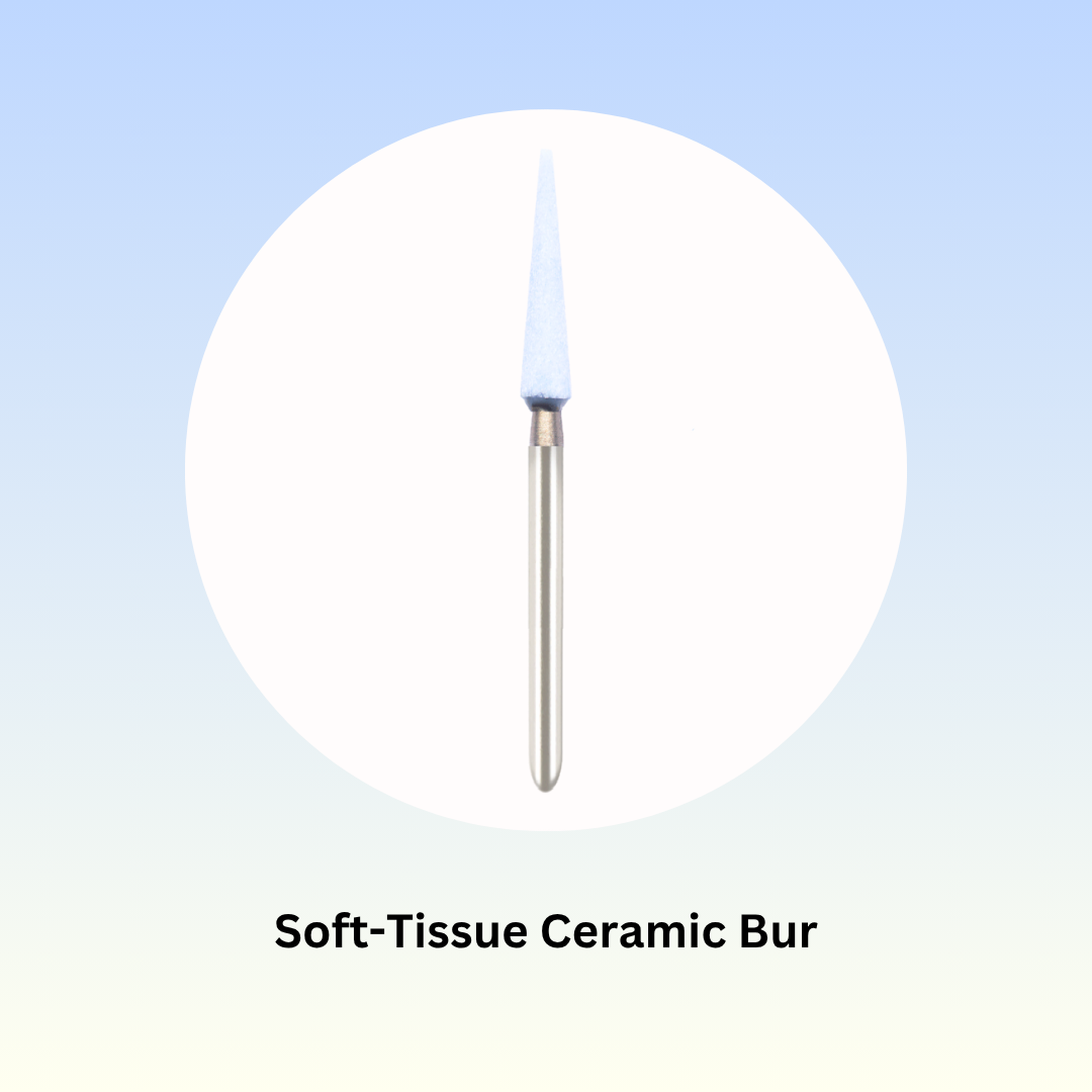
Soft tissue trimming and contouring play crucial roles in creating the ideal environment for graft placement. The use of specialized ceramic burs, such as Mr Bur Soft Tissue Trimming Ceramic Powder Bur FG (SOFT-T) and Mr Bur Ceramic Bur for Soft Tissue Trimming series (ST-1, ST-2 and ST-3), offers significant advantages over traditional cutting instruments.
-
These ceramic burs generate minimal heatduring operation, reducing the risk of thermal damage to delicate tissues and preserving cellular integrity, which is essential for optimal healing.
-
The precision offered by ceramic burs allows for controlled tissue removal and fine contouring, enabling the surgeon to create smooth, well-defined margins that support graft adaptation and stability.
-
The variety of shapes, ST-1, ST-2 and ST-3, offers tailored solutions for different clinical needs, from fine trimming to broader contouring.
-
Ceramic material retains sharpness longer than conventional burs, ensuring consistent cutting performance throughout the procedure.
Proper tissue preparation also involves:
-
Careful debridement of root surfaces
-
Removal of irregular soft tissue margins
-
Creation of a stable, well-vascularized recipient bed
Autogenous Graft Techniques
Autogenous soft tissue grafts remain the cornerstone of mucogingival surgery, particularly for root coverage and keratinized tissue augmentation. Among these, the connective tissue graft (CTG) is widely recognized as the gold standard, offering high predictability, long-term stability, and natural esthetic outcomes.
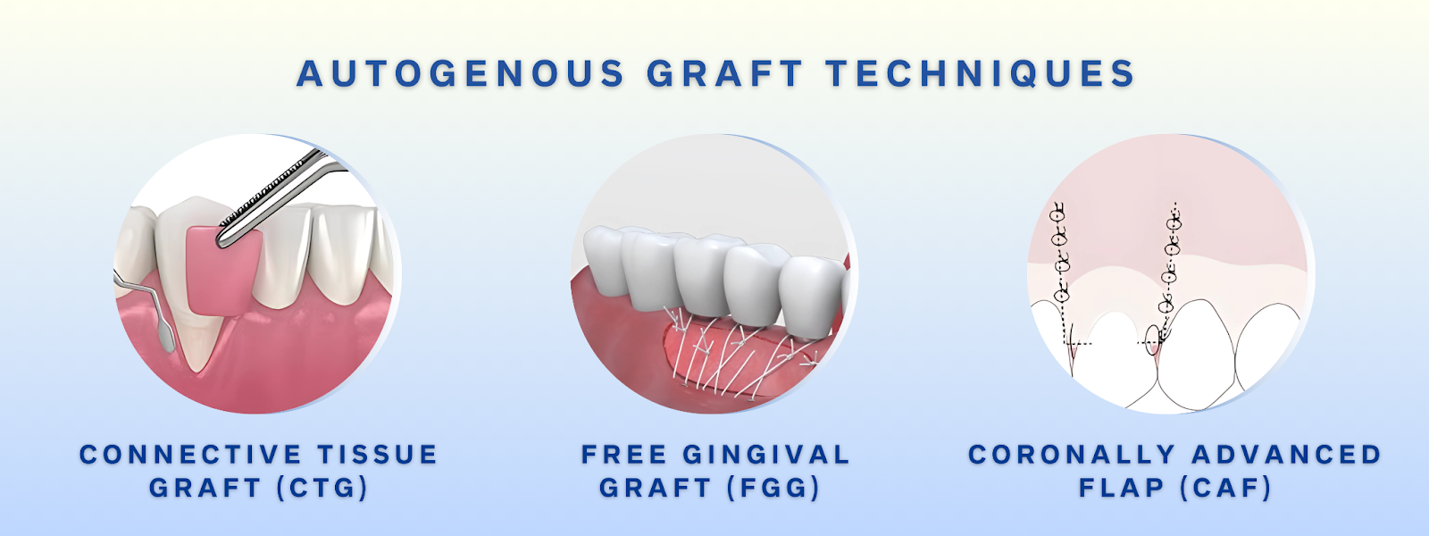
1. Connective Tissue Graft (CTG)
CTG success hinges on three core pillars: harvesting technique, graft thickness, and recipient site preparation.
-
Harvesting methods, such as the trap doorand single incision techniques, are chosen based on palatal anatomy and surgeon preference. The goal is to obtain a 1.5–2.0 mm thick graft while preserving donor site integrity.
-
Anatomical awareness is essential. Careful evaluation of the greater palatine artery and surrounding landmarks minimizes risk and enhances patient comfort.
-
Precision matters. Using sharp instrumentation for clean incisions improves healing and limits post-operative discomfort.
To optimize recipient site outcomes, clinicians must create a partial-thickness flap with sufficient coronal advancement potential. During this phase, the use of Mr. Bur Soft Tissue Trimming Ceramic Powder Bur FG and Ceramic Bur for Soft Tissue Trimming series burs allows for atraumatic epithelial removal, granulation tissue debridement, and marginal refinement without compromising vascular supply.
2. Free Gingival Graft (FGG)
While not typically used for root coverage, FGG remains the graft of choice for increasing the zone of keratinized tissue, especially in patients preparing for implants or managing mucogingival defects.
-
Graft success depends on donor site quality, color matching, and controlled thickness.
-
Mr. Bur ceramic burs provide precise recipient bed preparation, ensuring excellent tissue interface and graft stabilization.
3. Coronally Advanced Flap (CAF)
CAF can be used alone or in combination with CTG to treat Miller Class I and II or RT1 recession defects.
-
Achieving passive flap mobility and a stable coronal position is essential.
-
Releasing incisions and muscle insertions must be prepared carefully to preserve perfusion.
-
During this process, ceramic burs from Mr. Bur allow controlled removal of epithelial collars or scar tissue, improving adaptation while maintaining vascular support.
Alternative Grafting Materials
While connective tissue grafts (CTG) remain the gold standard for root coverage, alternative grafting materials offer excellent options when autografts aren't feasible due to patient preference, anatomical limitations, or morbidity concerns.
1. Acellular Dermal Matrices (ADM)
ADM grafts are derived from processed human tissue, designed to retain the natural collagen structure while eliminating immunogenic cells.
Advantages:
-
No second surgical site required
-
Unlimited and consistent supply
-
Supports revascularization and soft tissue integration
These are often paired with coronally advanced flaps for moderate to large gingival recession defects.
2. Xenogeneic Collagen Matrices
These collagen-based scaffolds, sourced from animal tissue, serve as biocompatible barriers and support tissue regeneration.
Best suited for:
-
Augmenting keratinized tissue
-
Covering shallow gingival recessions
-
Minimally invasive soft tissue procedures
While outcomes are less predictable than autografts, they are useful when minimal morbidity and faster recovery are priorities.
Advanced Surgical Techniques and Instrumentation
Modern soft tissue grafting techniques aim to enhance outcomes while minimizing patient trauma. The tunnel technique, which avoids vertical incisions, preserves vascular supply and reduces healing time, making it ideal for treating multiple adjacent recession defects.
Microsurgical approaches, using magnification and fine instruments, offer improved precision, smaller incisions, and faster recovery, though they require advanced training.
Specialized ceramic burs, such as those from Mr. Bur, play a key role in these techniques by enabling controlled soft tissue trimming with minimal heat generation, critical in preserving delicate tissues.
Many advanced protocols also incorporate regenerative materials, and their success often depends on precise tissue preparation, an area where Mr. Bur ceramic trimming burs excel.
Dental practitioners in the UK require tools that can handle the highest standards of care. Mr. Bur’s range of precision-engineered dental burs delivers the reliability and performance that UK dental practices demand. From London to Birmingham, our burs are trusted by dental professionals to provide exceptional outcomes, ensuring that every procedure is performed with the utmost accuracy.
Diamond Burs, Carbide Burs, Surgical & Lab Use Burs, Endodontic burs, IPR Kit, Crown Cutting Kit, Gingivectomy Kit, Root Planning Kit, Orthodontic Kit, Cosmetic Restorative 3-in-1 Kit FG, Composite Polishers, High Speed Burs, Low Speed Burs

